The first element in contact with the ground - in an anatomical position - it plays an essential role in balance, cushioning and propulsion.
The adult foot comprises 26 bones - that is, for both feet, a quarter of those making up the entire human skeleton -, 16 joints, 107 ligaments ensuring their protection and limiting mobility, 20 muscles which allow their movement.
The specialist doctors are podiatrists (Spain, England, Italy, Portugal, Canada and the United States).
|
|
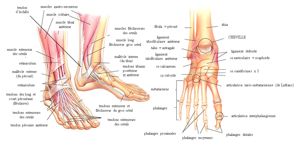
Hindfoot
The hindfoot is the posterior portion of the tarsus. It is made up of the talus and the calcaneus, the talus being above and slightly medial to the calcaneus, while being shorter than the latter. The talus is one of the ankle bones; it articulates above with the tibia mainly, and also with the fibula (or fibula). The talus also articulates downwardly with the underlying calcaneus, and its anterior end, located medially in the foot, articulates with one of the bones of the anterior row of the tarsus, the navicular bone. The calcaneus is the heel bone, and it is the largest bone in the foot. It articulates at the top with the talus, at the level of the anterior portion of its upper face. The posterior third of this same face receives the calcaneal tendon (or Achilean tendon) from the triceps sural muscle of the calf. The calcaneus is oriented forwards, upwards and laterally; its anterior end articulates with the cuboid bone. Midfoot The midfoot is the anterior part of the tarsus. It is made up of five bones. Medially there is a double row: these are the navicular bone posteriorly and the three cuneiform bones anteriorly. Laterally, there is only one bone: the cuboid bone. Concerning the medial part, the navicular bone therefore articulates: behind with the talus, laterally with the cuboid bone, in front with the medial, intermediate and lateral cuneiform bones. These occupy a relative position between them consistent with their names and are each in relation forward with one of the first three metatarsals. The lateral cuneiform bone is also articulated on its lateral surface with the cuboid bone. The lateral part of the midfoot is occupied by the cuboid bone, which therefore articulates: on its posterior surface with the calcaneus; on its medial surface, behind with the navicular bone and in front with the lateral cuneiform bone; on its anterior surface, with the fourth and fifth metatarsals. Forefoot The forefoot corresponds to the metatarsus and phalanges. The metatarsal is made up of the five metatarsals, similar to the metacarpal bone of the hand. The metatarsals are articulated at their base with: the bones of the distal row of the tarsus, in order the medial, intermediate and lateral cuneiform bones for the first three, and the cuboid bone for the last two; and with the base of the adjacent metatarsal (s). At the level of their head, distally, the metatarsals are each in relation to the base of each respective proximal phalanx. The phalanges of the foot are arranged in the same way as those of the hand; a proximal phalanx is articulated in front with a middle phalanx, itself articulated in front with a distal phalanx. As with the hand, the first phalanges are only two, the proximal and the distal. Finally, each metatarsal and the corresponding phalanges together form a single structure called a radius. Plantar surface The sole of the foot is a special region as it supports the weight of the body, which is mainly distributed over the posterior part of the calcaneus and the heads of the metatarsals. Arch of the foot The plantar surface is relatively hollow, which is due to the fact that the skeleton of the foot is organized in the form of an arch with a lower concavity. Three arches are thus described: longitudinal, medial and lateral, and transverse. The medial longitudinal arch is the most important in terms of length and curvature; it is made up, from back to front, of the calcaneus, the talus, the navicular bone, the cuneiform bones and the first three metatarsals. The lateral longitudinal arch is much less pronounced; it consists of the calcaneus, the cuboid bone and the last two metatarsals. The transverse arch consists of the cuneiform bones, the cuboid bone and the base of the metatarsals. This arching arrangement is made possible by the plantar fascia which is a band of dense connective tissue extending from the posterior part of the calcaneus to the heads of the metatarsals. The ligaments of the foot are also involved, as are the muscles and tendons of the plantar surface. |